Digital
Smile Design
Why Smile Design
Four complementary goals that structure the modern aesthetic approach and make it reproducible, case after case.
Clinical photography6
The objective, reproducible foundation of aesthetic diagnosis in dentistry.
Clinical photography forms the objective basis of any aesthetic analysis. It allows an in-depth reading of facial and dental characteristics, and serves as a communication medium between the clinician, the laboratory and the patient.
A standardized series (controlled distance, focal length and exposure) guarantees the reproducibility of the analysis and the reliability of the final design.
Video analysis complements photography by revealing the natural dynamics of the smile: phonation, lip animation, and spontaneous movements that still images alone cannot capture.
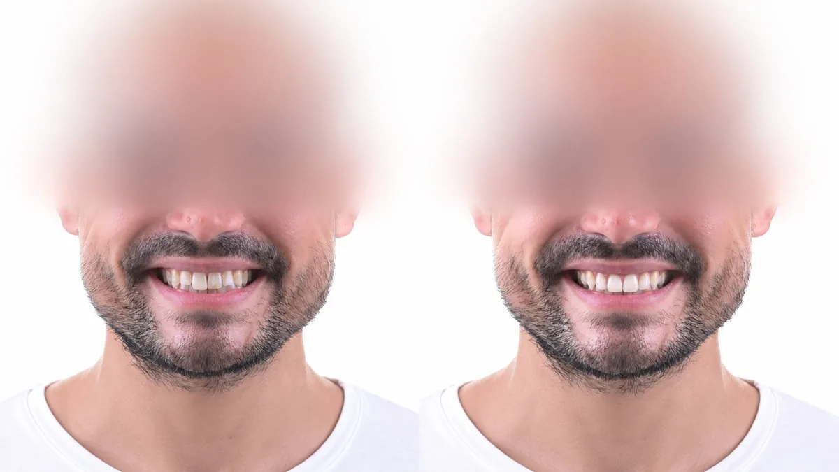
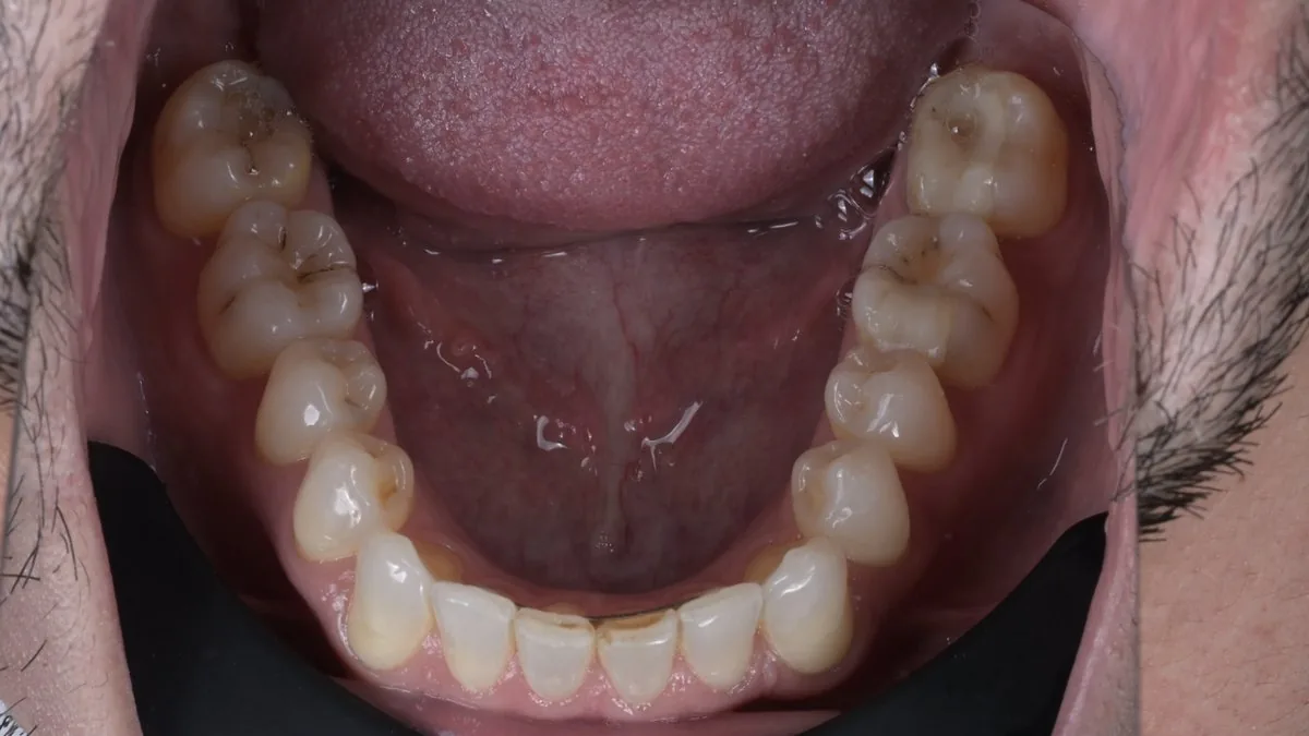
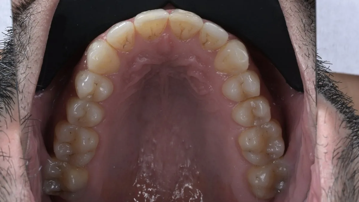
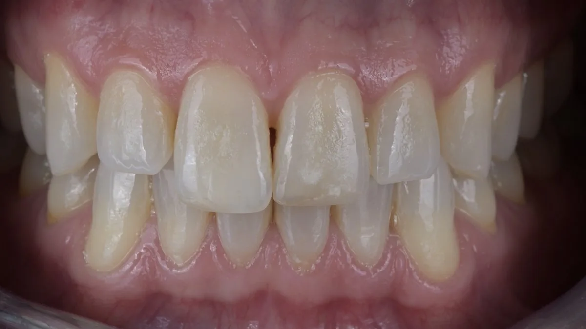
The three key photographs
The fundamental basis of aesthetic diagnosis and smile analysis: each one answers a specific clinical question.

- Assessment of the smile line
- Analysis of symmetry and the aesthetic plane
- Observation of lip dynamics
- Overall perception of the smile within the face
- Width-to-height proportions of the teeth
- Study of tooth axes and the midline
- Assessment of embrasures and contacts
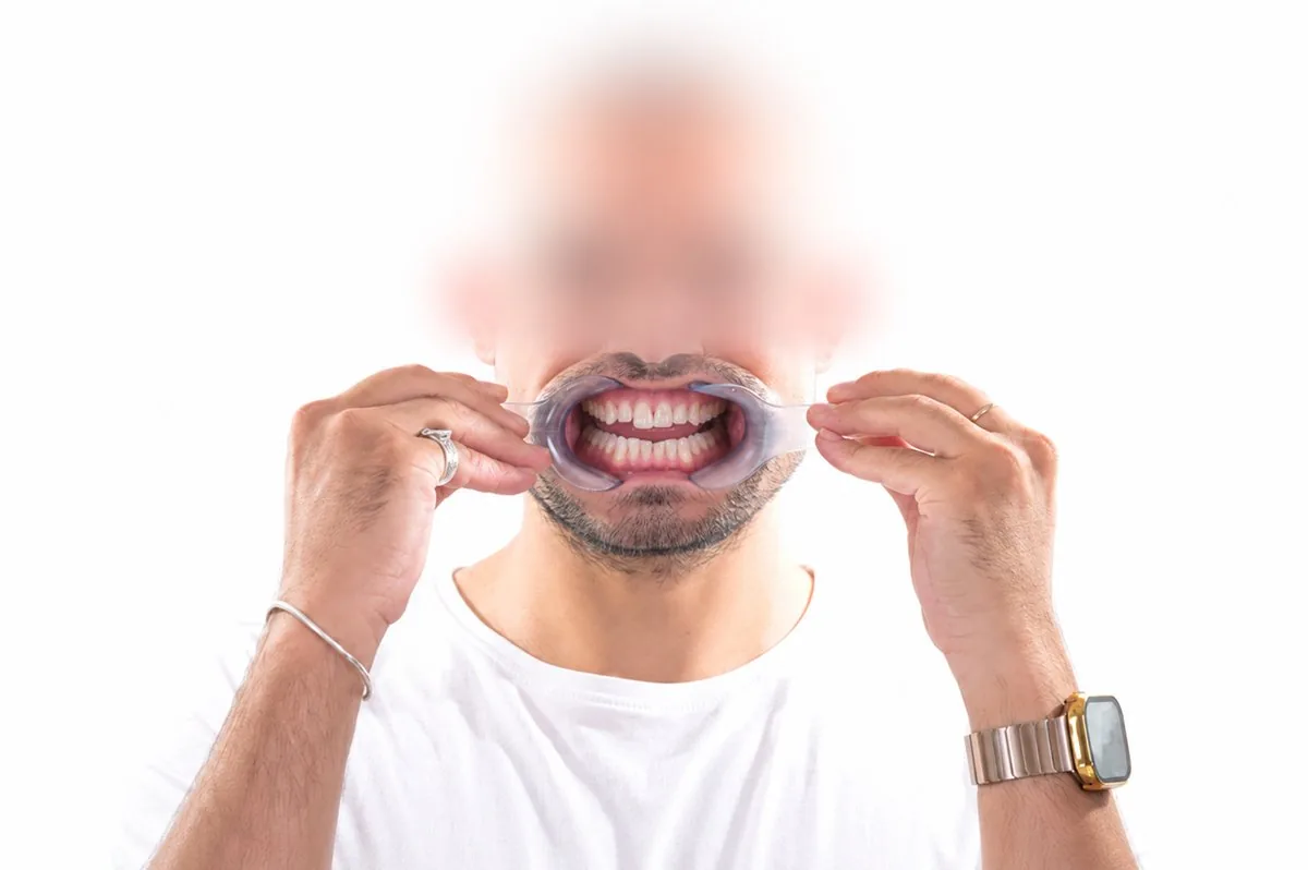
- Complete visualization of the arch

- Quantification of incisal display when smiling
- Assessment of the smile line and lip support
- Analysis of the tooth-lip relationship in animated posture
- Essential reference for anterior design
Advanced digital analysis4
DSD relies on a rigorous reading of facial landmarks to align the prosthetic project with the architecture of the face.
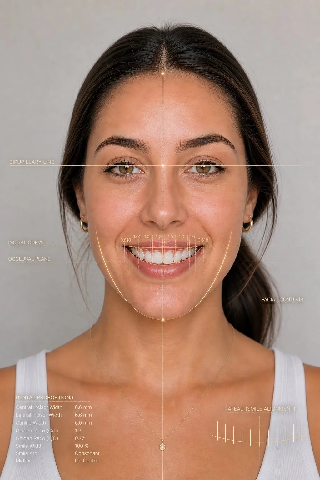
- Interpupillary line
- Midline
- Incisal curve
- Occlusal plane
- Facial shape
- Tooth proportions
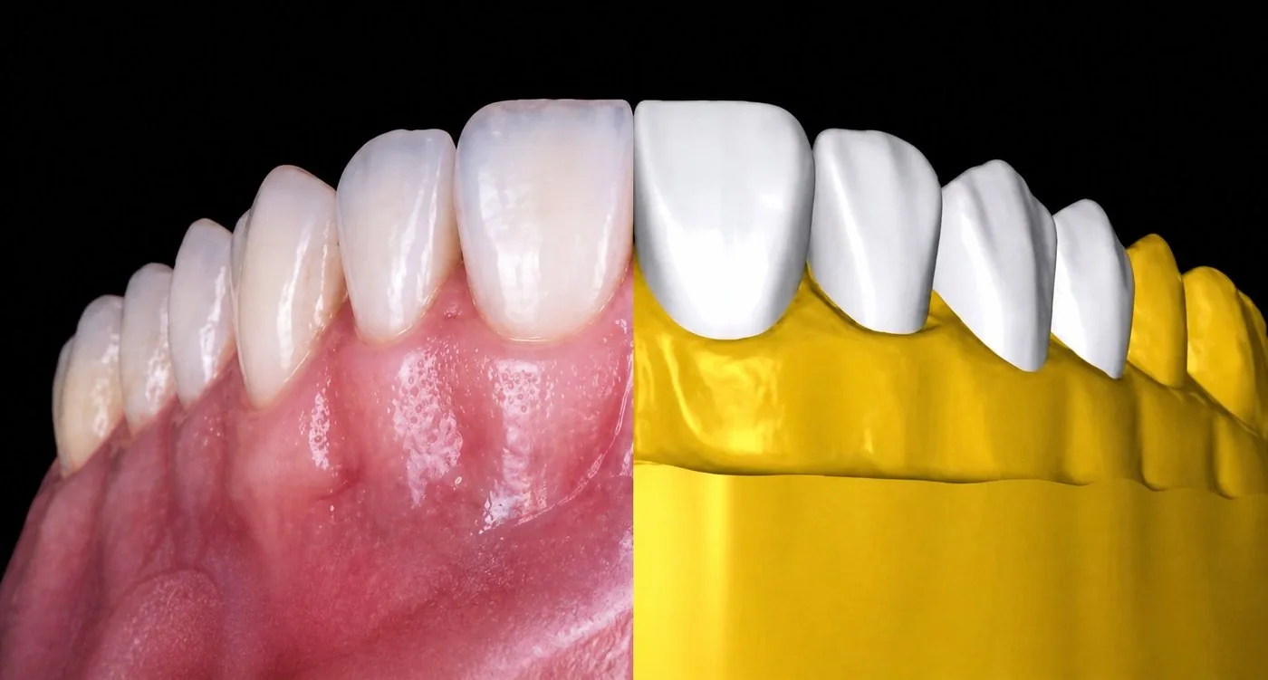
2D and 3D: two complementary approaches
An initial flat reading, then volumetric modeling. Each answers a different question: how they connect determines the result.
Initial visualization
on photographs
- Rapid identification of proportions
- Reading of aesthetic lines
- Simplified patient communication
- Validation of the project's direction
Volumetric modeling
under real conditions
- Adaptation to actual tooth volumes
- Surgical and prosthetic precision
- Validation under clinical conditions
- Direct transfer to 3D printing and the mock-up
The integrated clinical workflow7
Five steps, from photographic diagnosis to final patient validation.
Complementary photographs
Additional views that refine the design and allow advanced customization of the aesthetic project.

Three pillars, one method
What the photographic and digital approach concretely brings to each clinical case.
Scientific references
All methodological and clinical statements in this presentation are supported by recent peer-reviewed sources (PubMed). Each number N in the document points back here.
- Impact of artificial intelligence-based digital smile design on patient and clinician satisfaction and facial esthetic outcomes: A systematic review and meta-analysis. Digital Health. 2025;11. PMC12536214
- Digital Smile Design and Patient-Centered Outcomes in Esthetic Restorative Dentistry: A Systematic Review. Cureus. Published November 29, 2025. Cureus
- The Recent Use, Patient Satisfaction, and Advancement in Digital Smile Designing: A Systematic Review. Cureus. 2024;16(6):e62459. PMID 39022468
- Integrating digital smile design into restorative Dentistry: A narrative review of the applications and benefits. Saudi Dent J. 2024;36(4):561-567. PMID 38690398
- Assessment of Patient Satisfaction and Treatment Outcomes in Digital Smile Design vs. Conventional Smile Design: A Randomized Controlled Trial. J Pharm Bioallied Sci. 2024;16(Suppl 1):S669-S671. PMID 38595496
- Three-dimensional analysis of posed smile in adults: A scoping review. J Dent Sci. 2024;19(2):773-786. PMID 38618097
- Interdisciplinary guided dentistry, digital quality control, and the "copy-paste" concepts. J Esthet Restor Dent. 2021;33(7):988-995. PMID 33899323
- Parameters to Improve the Accuracy of Intraoral Scanners for Fabricating Tooth-Supported Restorations: A Review. J Esthet Restor Dent. 2025. DOI 10.1111/jerd.13364
Presentation document for educational purposes. The cited references are no substitute for reading the original sources in full or for case-by-case clinical evaluation.
Hugo Philippe FUSARO
Emailcontact@hpfdentalsolutions.com Phone+33 6 15 39 70 53 LinkedInView profile →